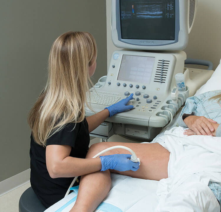
Vascular ultrasound is a non-invasive method which helps doctors to diagnose certain problems like deep vein thrombosis (DVT) or any other condition related to veins in the body. It is a way to see the blood flow and can be used to detect and assess abnormalities in the veins and arteries.
How it works?
It uses a high frequency sound waves that travel through the body. These waves pass through the soft tissues and are absorbed by the blood vessels and organs. The sounds that are received by the transducer help the doctor to visualize the blood flow. This imaging is usually done through the skin, but it can also be done through the ultrasound machine.
When you undergo this test, the doctor will use a gel-like substance to clean your skin before he presses the transducer against it. A machine will then take images of your vein or artery, and it will appear as a dark line on the screen. The area being scanned will appear lighter than the surrounding tissue.
This test is safe and painless and the doctor can perform it in a matter of minutes. The whole procedure takes only about 5 minutes, and after that, you can go back home. You will have to lie still for about an hour.
Why should you get it done?
Vascular ultrasound is used to detect problems related to the veins and arteries like DVT (deep vein thrombosis), leg ulcers, varicose veins, arterial disease, and other conditions. It helps doctors to detect and diagnose clots in the veins or arteries. It helps in the assessment of blood flow and can also help to determine if there is any damage to the vessel walls. This test is also used to help in the management of various heart conditions like coronary artery disease.
Vascular ultrasound is also used to assess the size of the veins and arteries. This test is used to identify the location of vein valves, which is helpful in preventing future DVT. This test is also used to evaluate the size of the veins, especially in the legs, for conditions such as varicose veins.
Vascular ultrasound is used to screen for deep vein thrombosis (DVT) or any other condition related to veins. It can help to detect blood clots, which are a major cause of blood clots in the veins.
How it is done?
Vascular ultrasound is usually done using an ultrasound machine. The doctor will use a gel-like substance to clean your skin before he presses the transducer against it. A machine will then take images of your vein or artery, and it will appear as a dark line on the screen. The area being scanned will appear lighter than the surrounding tissue.

The doctor will then use the ultrasound machine to create a 3D image of your veins. This will help him to diagnose any abnormality in the veins, such as varicose veins, blood clots, DVT, or leg ulcers. This test is also used to evaluate the size of the veins, especially in the legs, for conditions such as varicose veins.
Vascular ultrasound is used to assess the size of the veins, especially in the legs, for conditions such as varicose veins.
What are the risks involved?
Vascular ultrasound is safe and painless. There is no risk involved in this test and you can go back home after it. You will need to lie still for about an hour. You may feel some pressure on the area being scanned but it will not hurt. You should let your doctor know if you have had previous problems with venous thrombosis (blood clots).
You will need to be in good health to undergo this test. If you have any conditions like heart problems, diabetes, kidney problems, cancer, or high blood pressure, then you cannot undergo this test. Your doctor will give you a full medical history before performing this test.
What are the benefits?
This test helps doctors to detect and diagnose problems related to the veins and arteries. It helps in the assessment of blood flow and can also help to determine if there is any damage to the vessel walls. This test is also used to help in the management of various heart conditions like coronary artery


